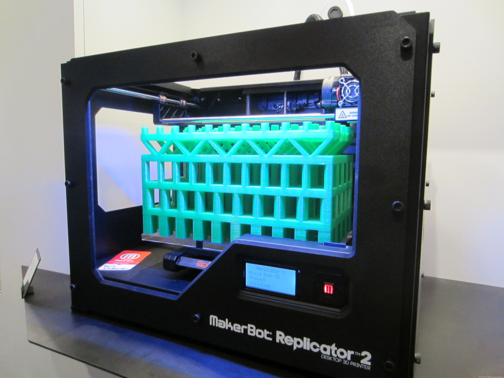
3D Printing technology is rapidly taking over healthcare and medical industries. Doctors like Anthony Atala are already seeing significant progress in utilizing the technology to produce tissues and organs with an unprecedented level of precision and accuracy.
Over the past few years, the global 3D printing market has evolved into a potent multi-trillion dollar market, with countries including Japan, the US and China leading the development of the technology. Increasing allocation of resources and capital into the development of 3D printing technology has led to the experimentation of various filaments.
Anthony Atala from the Wake Forest Institute for Regenerative Medicine in Winston, North Carolina, is relying on a 3D printing technology-based system called Integrated Tissue and Organ Printing System (ITOP) to 3D print tissues and organs by utilizing cells as the main filament or component of the 3D printer.
In the past, medical and healthcare industries focused on the commercialization and implementation of 3D printing technology to design accurate scans of patients and derive custom-built prosthetics or structures of particular organs to conduct practice operations prior to engaging in the actual surgery.
However, only a limited group of institutions and researchers have found success in utilizing 3D printing technology to create cells, organs and tissues that can be replaced with actual components in the human body. Atala aims to focus on the development of cell printing, to ensure that the healthcare industry finds more innovative and efficient methods of treating patients with highly complex and sophisticated operations.
The key component of the 3D printing method being tested by Atala is the process of laying real cells one layer at a time with an enterprise-grade 3D printer. Necessary data and information on the structure of cells or organs are received by the ITOP from CT or MRI scans, which are usually made readily available by surgeons.
“We take a very small piece of their tissue. We then start to expand those cells outside of the body. We use those cells to create new tissues and organs that we can then put back into the body. You’re laying cells one layer at a time. The material is deposited onto the surface to create a three dimensional structure,” said Atala.
One major difference between Atala’s ITOP and other systems tested in the past is that ITOP uses cells extracted from a specific patient. The utilization of cells from a specific individual eliminates the possibility of rejection from the body of the individual, allowing 3D printing-based operations to continue without any boundaries.
Already, Atala and the rest of his research team at the Wake Forest institute carried out tests on rodents. According to a study published by Atala, the rodents in the study successfully developed new tissues, bones, muscles and cartilages on top of the 3D printed cells.
“The incorporation of microchannels into the tissue constructs facilitates diffusion of nutrients to printed cells, thereby overcoming the diffusion limit of 100–200 μm for cell survival in engineered tissues. We demonstrate capabilities of the ITOP by fabricating mandible and calvarial bone, cartilage and skeletal muscle. Future development of the ITOP is being directed to the production of tissues for human applications and to the building of more complex tissues and solid organs,” the study read.
Image Via: KYM
If you liked this article, follow us on Twitter @themerklenews and make sure to subscribe to our newsletter to receive the latest bitcoin, cryptocurrency, and technology news.

